Origins Of Thigh Tendons / Posterior Thigh Muscles Hamstrings Anatomie Knie Anatomie Physiotherapie : Anterior inferior iliac spine insertion:. Its tendinous origin is extensive, arising from the top of the pelvis (iliac crest), most of the lumbar vertebrae, and several of the lower thoracic vertebrae. Many collagen fibres make up a fascicle. Abductor of thigh lateral rotator of thigh flexor of the leg at knee joint. Upper limb trauma programme physioplus courses should fulfil requirements for professional development. Tendons vary in size and are somewhat elastic.
The adductors, the lateral it inserts to the medial surface of the tibia, between the tendons of the sartorius (anteriorly) and the both gluteus medius and gluteus minimus, deep to gluteus maximus, share the same origin, insertion. Related online courses on physioplus. Tenocytes synthesize the collagen fibres that they surround. The semimembranosus, so called from its membranous tendon of origin, is situated at the back and medial side of the thigh. For example, a man with a 1 centimetre biceps tendon will have greater potential for muscle mass than a man with a longer.
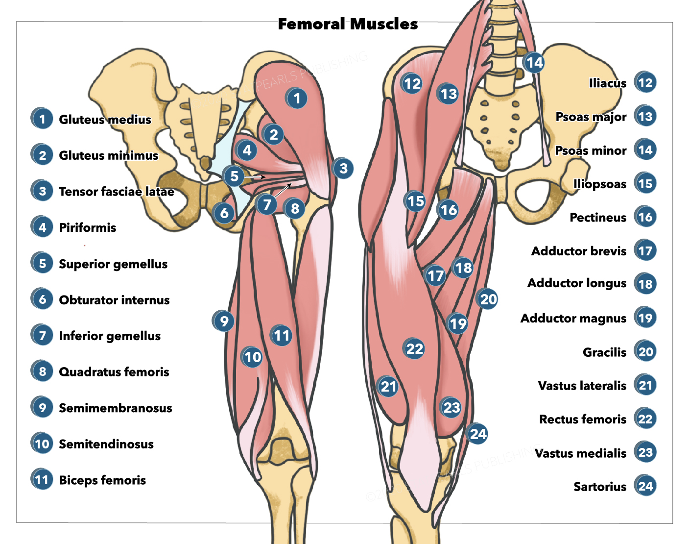
Anatomy Bony Pelvis And Lower Limb Femoral Muscles Article from www.statpearls.com Learn about their differences and the common tendons and ligaments commonly sustain injuries, which usually have similar symptoms and treatments. Attaches slightly distal to the gracilis and posterior to. Origin, insertion, and nerve supply of the muscles on the back of thigh. There are 3 tendons in the back of the thigh that connect the hamstring muscles to the ischial tuberosity (the sit bone) in the pelvis. The name gets its origin from its structure which is often conjoined or continuous. Quadriceps tendon to patella and then patellar ligament to tibial tuberosity action: Extends from the inner thigh bone to the lumbar vertebrae. Want to learn more about it?
Tendons are composed of bundles of collagen, predominantly type i, surrounding parallel rows of fibroblasts known as tenocytes.
When i buy whole chicken, it's usually pretty small birds, but even then the tendon is still quite big and makes it unpleasant to eat around. Tendons are similar to ligaments; It often occurs at its origin at the front of the hip. Medial shaft of the tibia just distal to the medial condyle; Patellar tendonitis (jumper's knee) is sometimes mistaken for quadriceps tendonitis due to the close working relationship within the soft tissues of the knee joint. Muscles, tendons, ligaments of the thigh… Learn this topic now at kenhub. Tendons attach muscle to bone. Anterior inferior iliac spine insertion: Quadriceps tendon to patella and then patellar ligament to tibial tuberosity action: All restaurants, chicken shops, etc. Becomes part of the quadriceps femoris tendon. The leg has one very thick tendon that finishes halfway down the bone, and the thigh has a few smaller tendons.
Medial aspect of the superior part of the ischial tuberosity via a shared tendon with the biceps femoris. It often occurs at its origin at the front of the hip. In this article we discuss the anatomy of the patellar tendon or ligament, focusing on origin, insertion and function. Tendons are tough, connective tissue that connects a skeletal muscle to a bone. Its tendinous origin is extensive, arising from the top of the pelvis (iliac crest), most of the lumbar vertebrae, and several of the lower thoracic vertebrae.
1 from The name gets its origin from its structure which is often conjoined or continuous. Tendons attach muscle to bone. Its tendinous origin is extensive, arising from the top of the pelvis (iliac crest), most of the lumbar vertebrae, and several of the lower thoracic vertebrae. Medial aspect of the superior part of the ischial tuberosity via a shared tendon with the biceps femoris. Attaches slightly distal to the gracilis and posterior to. Related online courses on physioplus. Gracilis, obturator externus, adductor brevis, adductor longus and adductor magnus. Causes leg flexion of the leg at the acetabulofemoral joint, extends leg at knee joint.
The thigh can be organized into five groups by the actions/location:
Because tendons receive less blood flow than muscle, they take a lot longer to respond to training than muscle. Tenocytes synthesize the collagen fibres that they surround. Superficial (middle) anterior thigh origin: Ligaments connect one bone to another, while tendons connect muscle to bone. Want to learn more about it? Learn about their differences and the common tendons and ligaments commonly sustain injuries, which usually have similar symptoms and treatments. The name gets its origin from its structure which is often conjoined or continuous. Quadriceps tendon to patella and then patellar ligament to tibial tuberosity action: Medial shaft of the tibia just distal to the medial condyle; Many collagen fibres make up a fascicle. For example, a man with a 1 centimetre biceps tendo. Tendons are tough, connective tissue that connects a skeletal muscle to a bone. Abductor of thigh lateral rotator of thigh flexor of the leg at knee joint.
Want to learn more about it? There are five muscles in this group; Learn about their differences and the common tendons and ligaments commonly sustain injuries, which usually have similar symptoms and treatments. Causes leg flexion of the leg at the acetabulofemoral joint, extends leg at knee joint. Superficial (middle) anterior thigh origin:
Leg Definition Bones Muscles Facts Britannica from cdn.britannica.com Tendons are tough, connective tissue that connects a skeletal muscle to a bone. Extends from the inner thigh bone to the lumbar vertebrae. Tendon length varies in all major groups and from monkey to person. Origin, insertion, and nerve supply of the muscles on the back of thigh. The adductors, the lateral it inserts to the medial surface of the tibia, between the tendons of the sartorius (anteriorly) and the both gluteus medius and gluteus minimus, deep to gluteus maximus, share the same origin, insertion. Related online courses on physioplus. The name gets its origin from its structure which is often conjoined or continuous. Anterior superior iliac spine insertion:
Extends from the inner thigh bone to the lumbar vertebrae.
The quadriceps tendon on top of the kneecap and the patellar tendon on the underside of it make up what is known as the quadriceps mechanism. A tendon or sinew is a tough band of fibrous connective tissue that connects muscle to bone and is capable of withstanding tension. For example, a man with a 1 centimetre biceps tendon will have greater potential for muscle mass than a man with a longer. Extends from the inner thigh bone to the lumbar vertebrae. Tendons attach muscle to bone. Anterior inferior iliac spine insertion: These have separate origins in the. It often occurs at its origin at the front of the hip. Related online courses on physioplus. In contrast, a ligament consists of bands of thick connective tissue that join bone to bone. Upper limb trauma programme physioplus courses should fulfil requirements for professional development. Tendons are similar to ligaments; Many collagen fibres make up a fascicle.